In a recent study published in Science Immunology, researchers explore gene expression profiles of distinct populations of intraepithelial lymphocytes (IELs), particularly T-cell IELs (T-IELs), within different regions of the gastrointestinal (GI) tract.
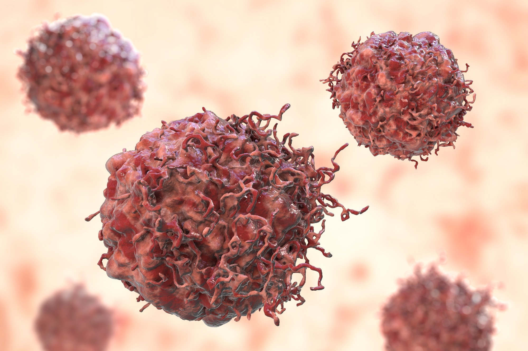
Study: TCF-1 limits intraepithelial lymphocyte antitumor Q1 immunity in colorectal carcinoma. Image Credit: Kateryna Kon / Shutterstock.com
What are T-IELs?
T-IELs constantly survey the epithelium of the GI tract to sense commensal microorganisms and signs of infection through T-cell receptors (TCR) or other receptors that help maintain GI tract barrier integrity.
Thus, T-IELs are critical in the communication network between the gut epithelium, microbiome, and diet. T-IELs are either thymic-derived, such as γδ and αβ T-cells, or induced, which develop from peripheral CD4+/CD8+ αβ T-cells.
About the study
In the present study, researchers performed single-cell ribonucleic acid sequencing (scRNA-seq) of mouse IELs to understand how distinctive populations of IELs in the GI tract perform different immune functions. To this end, over 13,400 IELs isolated from the stomach, small intestine, cecum, and colon of naïve C57BL/6 mice were analyzed.
Unsupervised clustering was used to investigate IEL diversity in the GI tract, especially T-cell clusters one, two, four, five, eight, and 10. Gene expression profiles of 200 up-and down-regulated genes in the colon and small intestine were also compared.
Flow cytometry (FC) was used to measure IELs and the factors they expressed to regulate T-cell differentiation in the small and large intestines. Notably, IELs play a crucial role in defense against colon cancer.
The human small intestine primarily absorbs nutrition from food; therefore, it encounters most dietary antigens. Comparatively, the colon absorbs fluids and comprises a higher microbial load and a more diverse microbial population.
Hypothesizing that colon-inhabiting microbiota could be responsible for driving T-cell factor-1 (TCF-1) expression, the researchers analyzed its expression in colon T-IELs in germ-free (GF) and specific pathogen-free (SPF) mice.
The Tcf7fl/flCD8αcre mouse model was used, in which the TCF-1 encoding TCF7 gene is knocked out from Cd8α expressing mature T-cells. Fluorescence-activated cell sorting (FACS) allowed the researchers to investigate the role of this gene in regulating colon T-IEL differentiation and function.
The MC-38 orthotopic murine model, in which MC-38 cells are implanted in the distal colon through colonoscopy, was also used to discern the role of the TCF7 gene and antitumor properties of γδ T-ΙΕLs. Recent studies have shown that Vγ7 and Vγ1 (γδ T-IEL subsets) are highly abundant in the mouse colon and help restrict tumor growth.
These findings were contextualized by analyzing the expression of IEL effector molecules in 63 samples from human patients with stage III colorectal cancer. To this end, tissue microarray methodology was used to assess the relative abundance of γδ T-cells in these samples.
Study findings
Stomach and small intestine IELs formed distinct clusters, whereas colon and cecal IELs essentially formed one cluster. CD3 Epsilon Subunit Of T-Cell Receptor Complex (CD3e) expression indicated that most IELs that originate in the small intestine, cecum, and colon were T-cells, whereas only a minor proportion of stomach-derived IELs were T-cells.
Major T-IEL subsets in the colon and small intestine were αβ and γδ T-IELs. Furthermore, γδ T-IELs exhibited notable increases in their cytotoxic phenotype compared with αβ T-IELs. Further analysis confirmed that stomach IELs were enriched in mast cells.
Previous studies have shown that heightened T-IEL defenses in draining lymph nodes from the small intestine compensate for deficiencies in adaptive T-cell responses to prevent infection and cancer and confer protection against dietary antigens. These findings demonstrate why different GI tract compartments have distinct T-IEL developmental requirements and functions.
Although similar IEL subsets were conserved across the small and large intestines, each region had a distinctive molecular profile. Colon T-IELs exhibited higher expression of TCF-1 but lower expression of effector and cytotoxic molecules.
All T-IEL subsets in the small and large intestines, particularly colon T-IELs, exhibited higher expression of nuclear receptor 77 (Nur77), a gene induced in T-cells upon TCR stimulation. Another notable difference between conventional T-cells and T-IELs was that colon T-IELs exhibited enhanced TCF-1 expression, which likely indicates that several factors regulate TCF-1 expression in T-IELs.
TCF-1 helped maintain the number of colon T-IELs over time, thus promoting self-renewal and the lifespan of conventional CD4+/CD8+ T-cell subsets.
Additionally, TCF-1 directly regulated the expression of genes in αβ and γδ T-IELs. Accordingly, it bound promoter regions of granzyme B (Gzmb), chemokine (C motif) ligand (XCL1), and tumor necrosis factor receptor superfamily (TNFRSF).
Some studies have shown that the intrinsic histone deacetylase (HDAC) activity of TCF-1 plays a crucial role in regulating T-IELs fate; however, further studies are needed to confirm these reports.
The absence of TCF-1 expression in human γδ T-IELs was associated with XCL1 and granzyme B expression. Since TCF-1 suppresses the antitumor properties of γδ T-IELs in humans, γδ T-IELs with enhanced antitumor properties could be used to develop immunotherapies using anti-cytotoxic T-lymphocyte-associated protein 4 (CTLA-4) to treat colorectal cancer, particularly its earlier stages.
Given the complex mechanisms of γδ T-IEL activation, additional research is needed to elucidate how γδ T cells could drive immunotherapy response in colorectal tumors lacking β2 microglobulin.
Journal reference:
- Yakou, M. H., Ghilas, S., Tran, K., et al. (2023). TCF-1 limits intraepithelial lymphocyte antitumor Q1 immunity in colorectal carcinoma. Science Immunology 8. doi:10.1126/sciimmunol.adf2163.
Credit: Source link


